
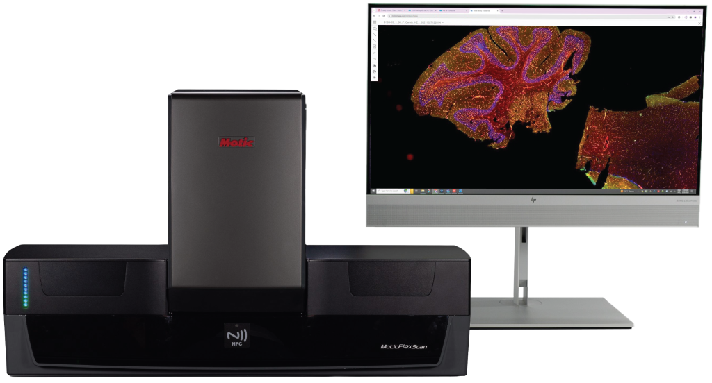
MoticFlexScan Pro 60
Elevate Your Research Efficiency with MoticFlexScan 60
Automate high-volume low-plex fluorescence whole slide imaging in an open-source tool tailored for any lab.
Accurate ROI Detection
The MoticFlexScan Pro 60 utilizes advanced tissue detection algorithms to accurately identify regions of interest, even in faintly stained samples. Its Preview Mode enhances scanning efficiency and streamlines your workflow for faster decision-making.
Ideal for H/E sample with IHC serial sections
Purpose-built for H&E and IHC applications, the system supports precise IHC quantification with optics designed for optimal visualization of staining patterns and cellular morphology—making analysis more reliable and consistent.
Enhance Camera Performance
Equipped with a 12-megapixel high-resolution camera, the FlexScan Pro 60 captures wide fields of view with exceptional clarity, allowing for the effortless observation of intricate histological details.
Rapid Focus Technology
For Kidney
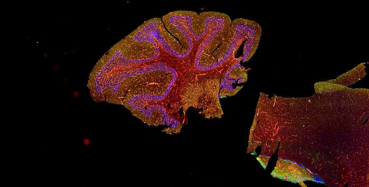
The MoticFlexScan 60 is a leading slide scanner for high-volume kidney tissue samples. It effortlessly handles kidney biopsy specimens stained with up to 10 different markers per case, efficiently scanning serial sections to enable thorough analysis of each digital slide. With support for 3-color, 2-color or single-plex FITC staining, it offers unparalleled flexibility. Its advanced imaging capabilities guarantee outstanding image quality, allowing for meticulous examination of crucial histological features. Furthermore, seamless integration with existing laboratory information systems and third-party image analysis software ensures smooth workflow, efficient data management, collaboration, and reporting.
For Skin
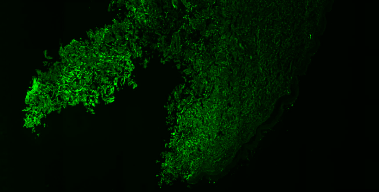
The MoticFlexScan 60 is the perfect digital pathology solution for dermatopathology labs. It handles diverse skin tissue samples, even challenging ones. Dermatologists can use two-plex staining protocols with DAPI as a counterstain for comprehensive visualization of epidermal layers, dermal features, and key histological elements. Its fluorescence overview Preview Mode feature optimizes scanning by helping users find fluorescently stained tissue or add focus points as needed. Additionally, it enables switching between brightfield and fluorescence imaging on any slides. This versatility allows dermatopathologists to leverage the most appropriate imaging modality for their specific diagnostic needs, whether it’s comprehensive visualization of histological structures or targeted analysis of fluorescent markers.